InMAGIC
The INMED Imaging facility (InMAGIC) offer to all users a large choice of microscopes and services. InMAGIC is under François Michel supervision and a scientific comity that include the institute director (Rosa COSSART) and two expert researcher (Agnès BAUDE and Valery Matarazzo).
Ressource reservation on OPEN Iris
Microscopy for biologistes
InMAGIC news

CircuitPhotonics
INMED and InMAGIC in collaboration with INT (Institut de neurosciences de la Timone) are laureates of EQUIPEX + 2020 with the project CIRCUITPHOTONICS, CENTER FOR IMAGING THE DYNAMICS OF NEURONAL CIRCUITS. With an investment of more than 4.5 million Euros in equipment, this new platform will be dedicated to the in vivo functional exploration of neuronal activity with the latest photonic microscopy techniques. Summary of the project
The technical offer include for types of microscope:
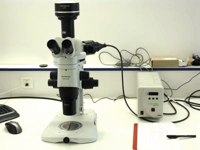
Stereomicroscopes
Two stereomicroscopes with camera are available, those give us the possibility to rapidly observe large samples in white transmission light or in epi-fluorescence mode with low magnification and large field of view.
(for more details)
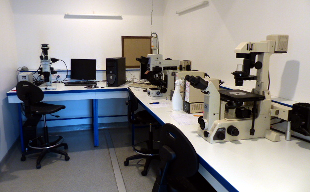
Wide-field Microscope
Three wide-field microscopes give the possibility to observe our samples with white transmitted light or fluorescence illumination. We offer both upright and inverted configurations with various magnifications (from 2 to 100 X), some are equipped with a camera and a motorized stage.

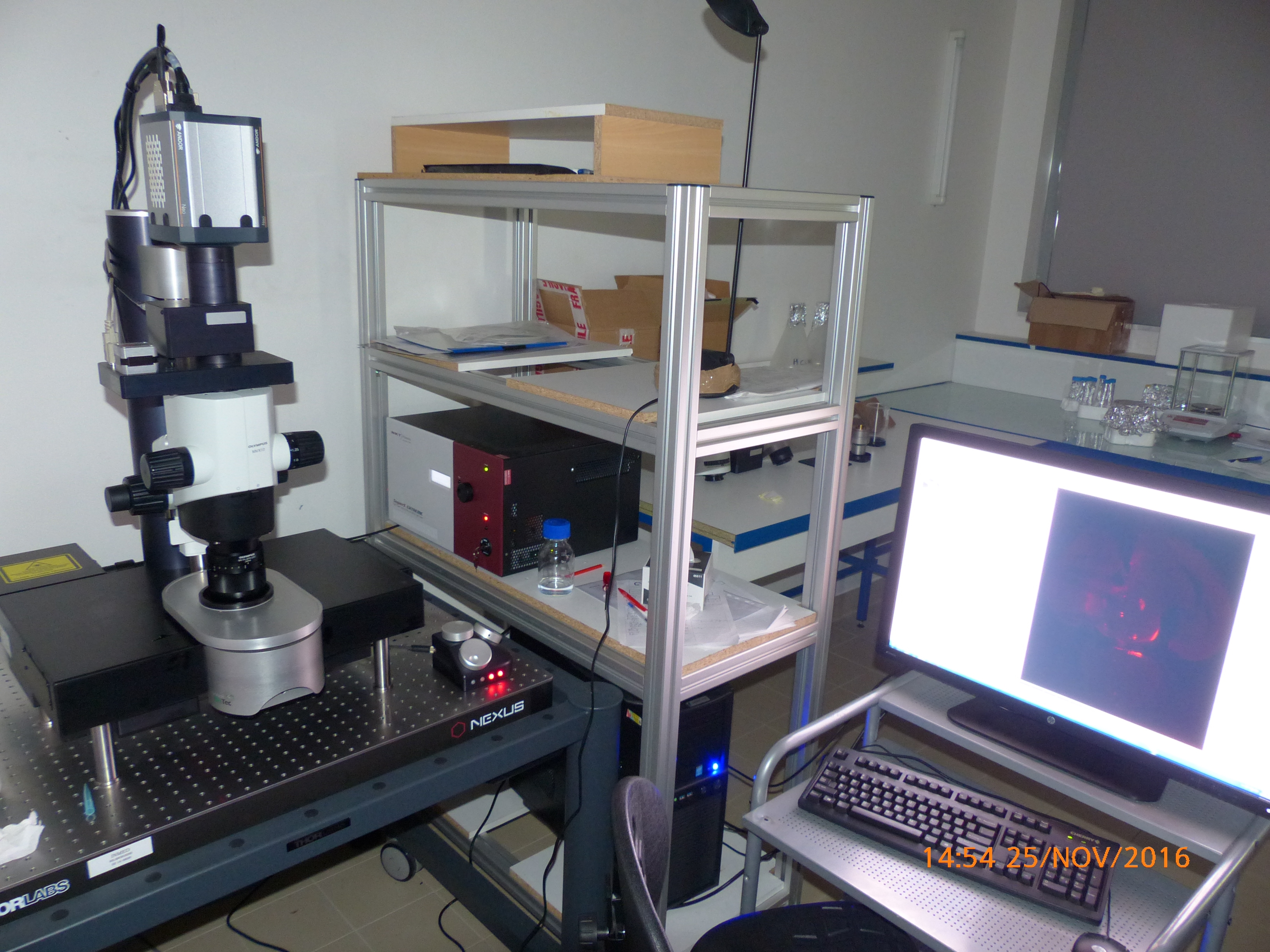
Confocal Microscope
Two confocal microscopes, technologically more complex, they offer the best of the photonic microscopy. They allow us to analyze and illustrate INMED experiments.
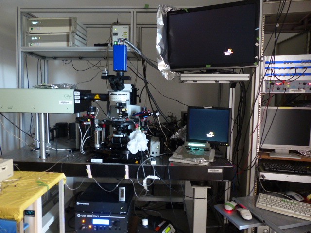
Multi-photon microscopes
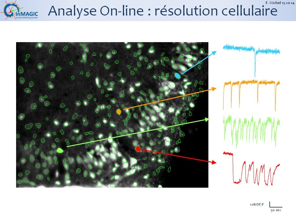
Our multiphoton microscopes are designed for dynamic imaging in samples kept alive and track in the same time electrical activity of neurons. It uses an advanced technology research that allows the feat to reconcile speed , resolution, large fields of observation and tissue preservation .
Light sheet microscope
Light sheet microscopes allows us to acquire large sample (as large as one cubic centimeter) with high resolution (les than 2 µm). It help us to follow large neuronal tracks in the whole brain or explore and discover new groups of specifically labeled cells far from the canonical and historical location for exemple.
An offer of practical and theoretical training (required for access to resources) and help with image analysis:
The platform also provides technical assistance for image analysis , access to specialized software and theoretical and practical training for a savvy use of all users
(see here for software and here for training )
An offer of advices and expermiental and technical developement
The platform engineers are also involved with the teams of the institute to conduct experiments and technical tune . In particular the Maturation of cortical GABAergic microcircuits team has two multiphoton microscopes : one for in vitro with optogenetic device and one for in vivo experiments .
Inmagic rules
Focus in InMAGIC

In a special issue of Sciences et vie junior (n°102) “light super power” François Michel speak about fluorescence microscopy power.
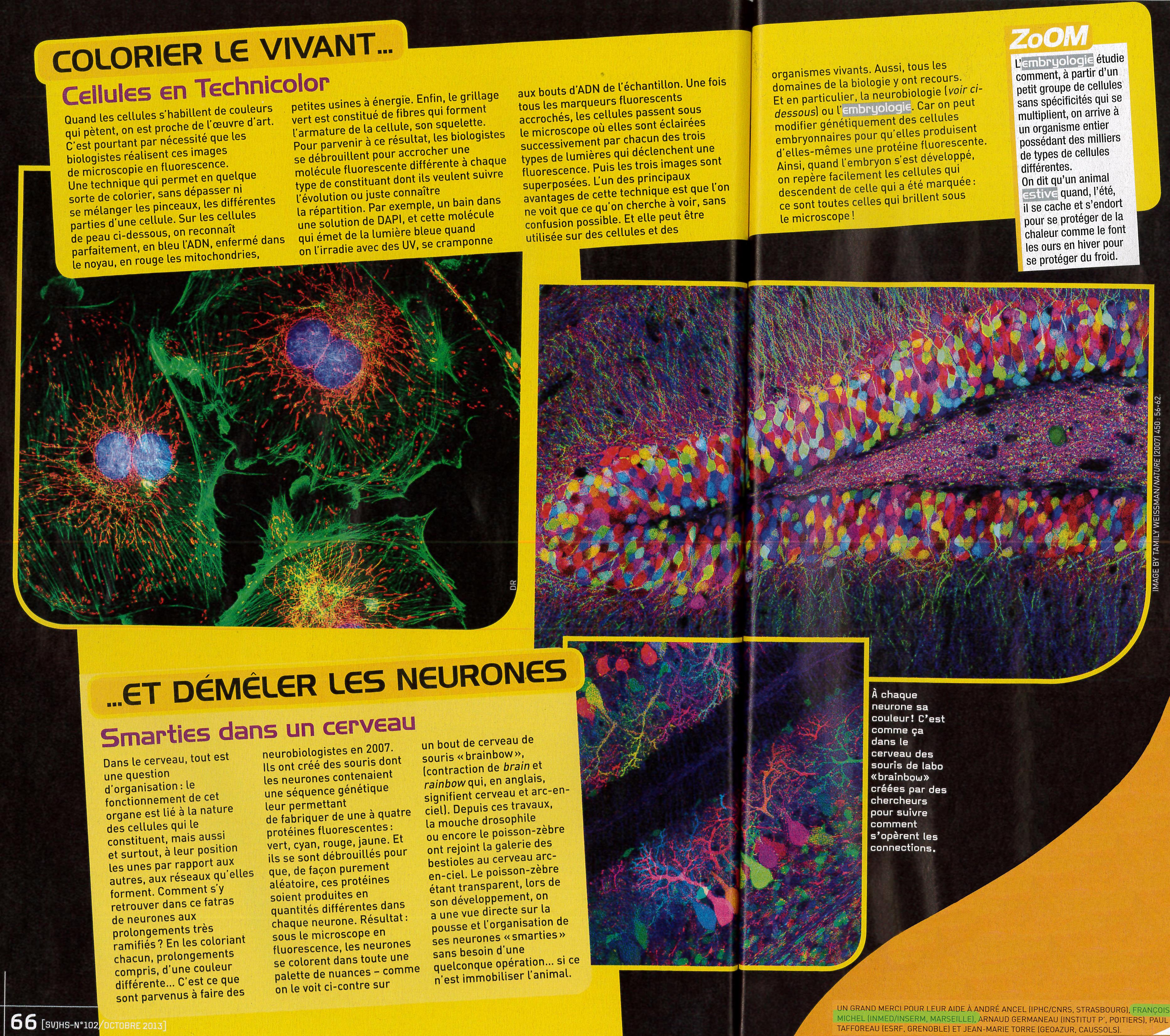
Let’s read: neurons as colored candies
A focus of a Brain paper involving François MICHEL in the letter of INSERM
