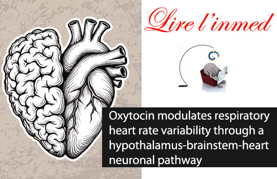
Brain’s heart beat. Without us noticing, our heart rate increases when we breathe in and decreases when we breathe out. This subtle modulation is thought to help the heart use its energy most effectively when the blood is freshly oxygenated. Emotional states alter this modulation, which is increased by oxytocin, a key agent of positive feelings. Here, the authors precisely mapped the associated circuits, from the oxytocinergic neurons in the hypothalamus, through the glycinergic neurons in the pre-Bötzinger complex, to the nucleus ambiguus and its neurons that innervate the heart. This circuit is activated during calming behavior. Their findings bring the heart and the brain ‘a beat’ closer together.
Scientific abstract: The variation in heart rate in phase with breathing is called respiratory heart rate variability (RespHRV). Relaxation and positive socio-emotional states can amplify RespHRV, yet the underlying mechanism remains largely unknown. Here we identify a hypothalamus–brainstem neuronal pathway in rodents through which oxytocin (OT) amplifies RespHRV during calming behavior. OT neurons from the caudal paraventricular nucleus in the hypothalamus regulate the activity of a subgroup of inhibitory neurons in the pre-Bötzinger complex, the brainstem nucleus that generates the inspiratory rhythm. Specifically, OT enhances the glycinergic input from OT-receptor-expressing neurons in the pre-Bötzinger complex to cardiac-innervating parasympathetic neurons in the nucleus ambiguus during inspiration. This leads to amplified respiratory modulation of parasympathetic activity to the heart, thereby enhancing RespHRV. We show that OT neurons participate in the restoration of RespHRV amplitude during recovery from stress in mice, indicating that OT acts centrally to regulate cardiac activity during a calming behavior.
The authors: J Buron, A Linossier, C Gestreau, F Schaller, R Tyzio, MS Felix, V Matarazzo, M Thoby-Brisson, F Muscatelli & C Menuet
Paru dans Nature Neusocience 2025